

Stress Fractures
Foot and ankle stress fractures are tiny cracks in the bone caused by overuse or repetitive stress, commonly seen in athletes and those who suddenly increase activity levels.
You had surgery. The hardware is in place. Your surgeon reviewed the imaging and told you the repair looks good. You are walking, which is more than you could do six months ago. And yet the pain and stiffness are significantly worse than you expected them to be at this point, the foot does not move the way it used to, and nobody has prepared you for what the next twelve months are going to look like.
This is one of the most common experiences in calcaneal fracture recovery, and it is almost never adequately explained to patients before or after surgery.
A calcaneal fracture — a fracture of the heel bone, most often caused by a fall from height or a high-energy impact — is among the most functionally complex injuries in foot and ankle medicine. The recovery timeline is longer than almost any other foot fracture, the long-term consequences are more variable, and the most significant complication is one that develops not immediately after surgery but in the months and years that follow, often when the patient assumed the hard part was already over.
Understanding what is actually happening inside the joint, why it is happening, and what can be done about it as it evolves is what this post is for.
What a Calcaneal Fracture Actually Does to the Joint
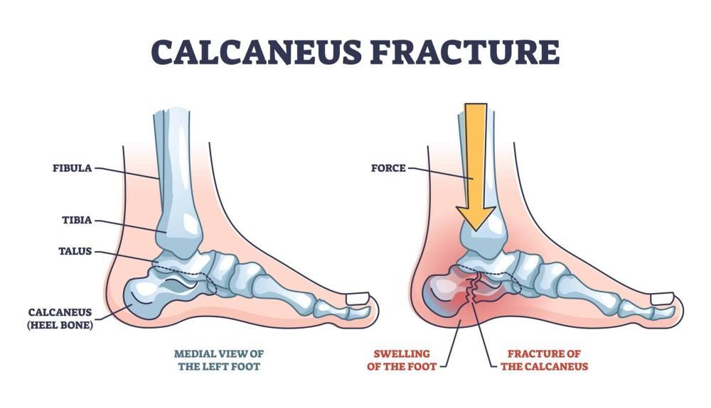
The calcaneus is the largest bone in the foot. Its primary function is structural: it forms the foundation of the hindfoot, transmits body weight from the leg to the ground, and forms the lower half of the subtalar joint — the joint that controls inversion and eversion of the heel and allows the foot to adapt to uneven terrain.
When the calcaneus fractures under high-energy impact, the force typically drives the talus downward into the top of the calcaneus, which collapses and shatters the joint surface. The articular cartilage — the smooth, load-bearing surface that lines the subtalar joint — is damaged at the moment of impact. That damage is immediate and irreversible. No matter how precisely the bone is reduced and fixed, the cartilage injury that occurred when the fracture happened cannot be surgically corrected. The hardware restores the architecture of the bone. It does not restore the joint surface to its pre-injury state.
This distinction matters enormously for understanding what recovery actually looks like, and why a technically successful surgery does not guarantee a pain-free outcome.
The severity of the initial joint surface damage is classified by the Sanders classification system, which uses CT imaging to categorize calcaneal fractures by how many pieces the joint surface has been broken into and how displaced those pieces are. Sanders type II fractures involve a single fracture line through the joint surface. Sanders type III fractures involve two fracture lines and three fragments. Sanders type IV fractures are severely comminuted — the joint surface is shattered into multiple pieces. The higher the Sanders type, the greater the initial cartilage damage, the harder it is to achieve an adequate reduction, and the higher the probability of significant post-traumatic arthritis developing over time. Long-term follow-up data shows that Sanders type III fractures are approximately four times more likely to require eventual subtalar fusion than type II fractures.
What Post-Traumatic Subtalar Arthritis Is and When It Develops
Post-traumatic subtalar arthritis is the deterioration of the subtalar joint cartilage following injury, driven by the initial damage at fracture, the altered joint mechanics that follow even a well-reduced repair, and in some cases by residual incongruity in the joint surface that could not be fully corrected surgically.
It does not develop overnight and it does not always develop to the same degree. But it develops in a substantial proportion of patients who sustain displaced intra-articular calcaneal fractures regardless of the quality of their surgical repair, because it is a consequence of the cartilage injury at the moment of impact rather than a consequence of anything the surgeon did or did not do correctly.
The typical timeline looks like this.
In the first six months after surgery, the dominant symptoms are related to bone healing, soft tissue recovery, and the gradual restoration of weight-bearing. The subtalar joint is stiff, which is expected. Pain during this period is largely mechanical — from the healing bone, the hardware, and the tissue that was disrupted during surgery.
Between six and twelve months, most patients are walking without support. Swelling may still fluctuate, particularly after prolonged activity. Calf strength is often significantly diminished after months of reduced weight-bearing and requires active rehabilitation. Nerve symptoms — numbness or tingling along the outer heel — are common and usually improve during this period, though not always completely.
Between twelve and twenty-four months, the bone remodeling is complete and the hardware is no longer doing active work. This is the window in which post-traumatic subtalar arthritis most commonly becomes symptomatic for patients who are going to develop it. The presentation is typically deep aching in the hindfoot after prolonged standing or walking, stiffness that is most pronounced in the morning and after periods of inactivity, difficulty on uneven ground, stairs, and slopes, and a progressive reduction in how long the patient can be on their feet before pain forces them to rest.
This is also the period during which patients who were not told to expect this experience the most anxiety, because they assumed that twelve months out from surgery meant they should be approaching normal — not developing new symptoms that feel like they are getting worse rather than better.
The Functional Questions Most Patients Are Not Getting Answered
The three questions patients most commonly carry into follow-up appointments — and most commonly leave without a clear answer — are about return to work, walking on uneven ground, and impact activities.
Return to work depends heavily on what the work involves. Desk workers and those in sedentary roles typically return within three to six months of surgery. Physically demanding work — construction, manual labor, prolonged standing on hard surfaces — typically takes twelve to eighteen months, and in some patients with significant joint damage it may require permanent modification of physical demands or a permanent change in role. Nonoperative management has been associated with up to six times the rate of late subtalar fusion compared to operative repair, which reflects both the importance of the initial treatment and the reality that returning to heavy physical demands on a compromised joint accelerates arthritis progression.
Walking on uneven ground is one of the most consistently reported long-term limitations after calcaneal fracture, even in patients with good surgical outcomes. The subtalar joint is what allows the foot to accommodate slope, gravel, grass, and irregular surfaces. When that joint is stiff — whether from the fracture itself, the repair, or developing arthritis — the foot cannot adapt the way it used to, and the ankle and knee compensate in ways that produce fatigue and secondary pain patterns. This limitation often persists for the full two-year recovery window and may remain in some degree permanently in patients with significant joint damage.
Impact activities — running, jumping, sports involving cutting and direction changes — are the last to return and may not return to pre-injury levels in patients with moderate to severe initial joint surface damage. The timeline for return to sports in appropriately selected patients is twelve to eighteen months from surgery at the earliest, with the caveat that developing subtalar arthritis changes that calculus significantly.
Management as the Joint Evolves
The management of post-traumatic subtalar arthritis after calcaneal fracture follows a spectrum, and where a patient sits on that spectrum at any given point determines what the appropriate next step is.
For patients with early or mild arthritis — hindfoot aching that is manageable with activity modification, supportive footwear and custom orthotics can meaningfully reduce load across the subtalar joint and extend the period during which the joint functions adequately without further intervention. Rigid or semi-rigid orthotic designs that limit subtalar motion are specifically effective for this purpose. Bracing options including hinged ankle-foot orthoses provide additional stability and load reduction for patients with more significant symptoms.
Corticosteroid injections into the subtalar joint offer targeted anti-inflammatory relief for patients with acute flares or periods of increased activity. They are not a long-term solution and have diminishing returns with repeated use, but they are a legitimate part of managing a joint that is progressing slowly and the patient wants to manage conservatively as long as possible.
For patients with end-stage symptomatic subtalar arthritis — pain that is no longer manageable with conservative measures, significantly limiting daily function, and not responding to injections — subtalar arthrodesis (fusion of the subtalar joint) is the definitive treatment. The procedure eliminates motion at the arthritic joint, which resolves the pain generated by that motion, at the cost of the residual subtalar range of motion the patient still has. Subtalar fusion for post-traumatic arthritis following calcaneal fracture produces good to excellent outcomes in 70 to 85% of patients, with meaningful improvement in pain and the ability to stand and walk on the affected limb. Patients who develop arthritis after a fracture that was well-reduced with restored calcaneal morphology tend to have better fusion outcomes than patients who develop arthritis after malunion from nonoperative management, which is one reason the quality of the initial repair has long-term relevance beyond the immediate post-operative period.
Increasingly, arthroscopic and minimally invasive subtalar fusion techniques offer the same fusion outcomes with smaller incisions, reduced soft tissue disruption, and in some series a faster return to weight-bearing — reflecting the same evolution toward less invasive surgery that has characterized other areas of foot and ankle reconstruction.
Why the Follow-Up Relationship Matters More Than Most Patients Realize
A calcaneal fracture is not a condition that is resolved at the point of hardware healing. It is a condition that sets a trajectory — for the joint, for the patient's function, and for the management decisions that will need to be made as the joint evolves over the next several years.
A follow-up relationship with a specialist who understands where the patient's joint currently sits on that trajectory, what the imaging shows about how it is changing, and what intervention is appropriate at each stage is the single most important factor in managing that trajectory well. The patient who is seen at six weeks and twelve weeks and told the hardware looks good and then not seen again until they are in severe pain at eighteen months has lost time that could have been used to intervene earlier, slow progression with conservative measures, and plan any eventual surgical intervention on an elective timeline rather than an urgent one.
The pain and stiffness you are experiencing at six or twelve months after a calcaneal fracture are not signs that something went wrong with your surgery. They are the documented natural history of a joint that was severely injured. What matters now is understanding where your joint is in that trajectory, what is coming next, and what options are available to manage it — because those options are most effective when they are applied early and adjusted as the joint changes rather than introduced only when the situation has already become severe.
If you have had a calcaneal fracture and feel like the explanation of what comes next has been incomplete, that conversation is exactly what we are here for.
Contact the Center for Foot and Ankle Restoration to schedule an evaluation.


